Understanding Dermatoscopy
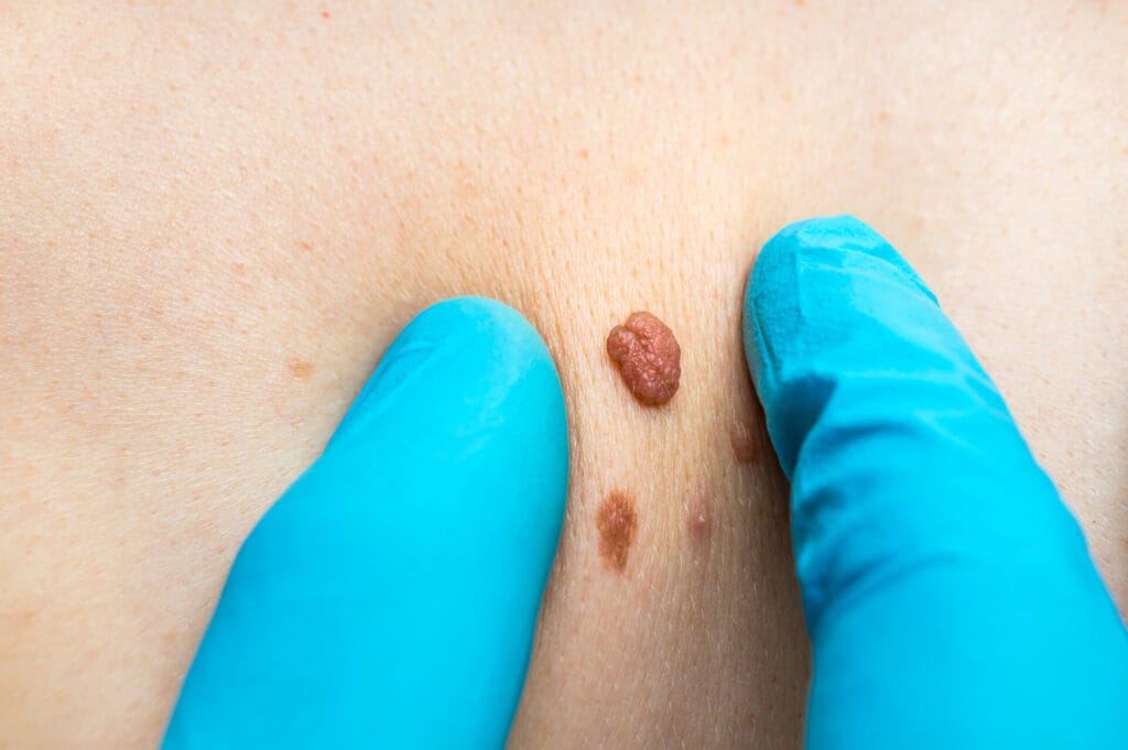
Dermatoscopy: Help us to detect any irregular mole/spot
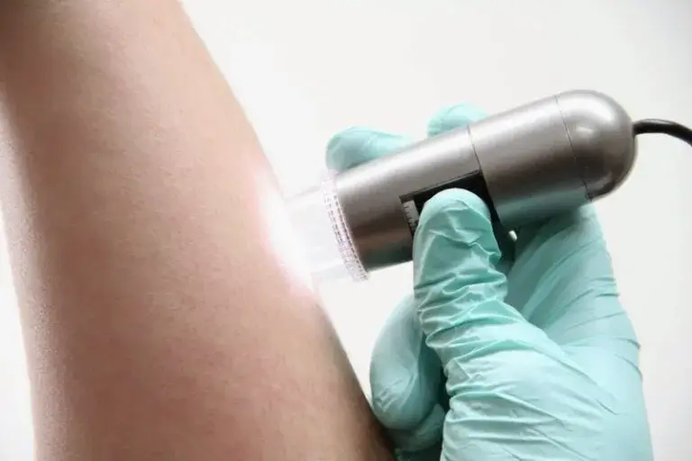
Dermoscopy is a non-invasive imaging technique used by us to examine skin lesions and moles. It involves the use of a handheld device called a dermatoscope, which provides a magnified and illuminated view of the skin’s surface.
Why It’s Useful:
Dermoscopy enhances the ability to detect skin cancer and irregular moles by allowing us to observe specific features not visible to the naked eye. These features include pigment patterns, vascular structures, and other subtle characteristics that can indicate the presence of skin cancer or abnormal growth.
Benefits:
- Early Detection: Dermoscopy enables early detection of skin cancer by identifying suspicious features in lesions or moles that may not be apparent during a visual examination alone.
- Improved Accuracy: By magnifying and illuminating the skin, dermoscopy enhances the accuracy of skin cancer diagnosis, reducing the likelihood of missed or misdiagnosed lesions.
- Guided Biopsies: Dermoscopy helps guide dermatologists in deciding which lesions require further evaluation through biopsy, ensuring that only suspicious or high-risk lesions undergo invasive procedures.